Dental X-rays are digital pictures of your teeth and jaws. Dentists use them to see parts inside your mouth that cannot be seen during a normal checkup. It is an X-ray that we see in general medicine for our chest, bones, or head. Dentists take a picture of the jawbone, nerves, sinuses, and tooth roots at an X-ray machine, called OPG.
What can dental radiographs detect?
Dental X-rays help your dentist find many oral health problems, including:
- Cavities, especially between teeth
- Decay under old fillings
- Bone loss in your jaw
- Teeth that have not come out or are stuck
- Abscesses (infections at the tooth root or between the gums and teeth)
- Cysts and some tumors
Dentists also use X-rays to decide if you are suitable for treatments such as implants, root canal, braces, or dentures. X-rays also help monitor healing after treatments like dental implants, bone grafts, or root canals.
Types of dental X-rays
There are two main types:
Intraoral X-rays
The sensor or film is placed inside your mouth. These X-rays help your dentist see details inside your mouth:
- Bitewing: Shows cavities between teeth and below the gum line.
- Periapical: Finds gum disease, bone loss, and cavities near tooth roots.
- Occlusal: Shows problems under your tongue or on the roof of your mouth, such as stuck teeth or broken jaw bones.
Extraoral X-rays
These pictures are taken with the film or sensor outside your mouth:
- Panoramic: OPG shows your whole mouth, including teeth, jaws, bones, nerves, and sinuses.
- Cephalometric: A side picture of your head, often used for planning braces.
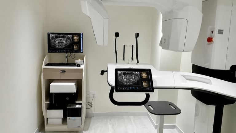
- Cone beam CT scan: CBCT gives 3D pictures of your teeth, jaws, nerves, and sinuses. It is often used for planning dental implants.
How do dental radiographs work?
Dental X-rays use a very small amount of radiation to take pictures of your mouth. The radiation beam passes through soft tissues and creates pictures of your teeth and bones. X-rays can be traditional film or digital imaging. Modern X-rays use 80% to 90% less radiation compared to traditional ones.
Early Detection of Dental Problems
Dental X-rays allow dentists to find possible problems that may not be seen during a regular visual check. This includes cavities between teeth, decay under existing dental work, and problems with the jawbone. Finding these issues early allows quick treatment and helps stop dental problems from becoming worse.
Assessment of Tooth and Jaw Development
X-rays are especially useful for checking the growth of teeth and jaws in children and teenagers. Dentists can identify problems such as improper tooth eruption, impacted teeth, or unusual jaw growth. This early information helps create the right treatment plan and supports proper oral development.
Evaluation of Bone Health
Dental X-rays give a clear picture of the jawbone and help dentists check its health and strength. This is important for many dental treatments, such as placing implants, where a strong and healthy jawbone is needed for long-term success.
Diagnosis of Periodontal Diseases
Periodontal diseases affect the gums and the tissues that support the teeth and can be difficult to diagnose without X-rays. These pictures show the amount of bone loss and help dentists create treatment plans for conditions such as gingivitis and periodontitis.
Treatment Planning and Monitoring
For many dental treatments, including braces, root canals, and tooth removal, X-rays play an important role in planning treatment and checking progress. They provide a complete view of the mouth structures. It helps dentists provide accurate and effective care, including procedures related to laser dentistry in Des Moines.
How are dental X-rays done?
Before taking dental X-rays, a technician may place a lead apron over your chest and a thyroid collar around your neck. This helps protect you from extra radiation. You will sit or stand in front of the machine while the technician places the film or sensor and takes the picture. Stay as still as possible.
Preparing for dental X-rays
Dental X-rays need no special preparation. The only thing you should do is brush your teeth before your appointment. This creates a cleaner environment for the people working inside your mouth. X-rays are usually taken before cleanings.
At the dentist’s office, you will sit in a chair with a lead vest across your chest and lap. The X-ray machine is placed beside your head to take pictures of your mouth. Some dental offices have a separate room for X-rays, while others take them in the same room where cleanings and other treatments are performed.
Are dental X-rays safe?
Dental X-rays are extremely safe as they use very low amounts of radiation. However, repeated exposure to high amounts of radiation can be harmful and may increase the risk of cancer. That is why dentists only take X-rays when needed. Your dentist will help you understand the risks and benefits.
How frequently is it safe to get dental radiographs?
If your teeth and gums are healthy, you may only need dental X-rays every six to eighteen months. But if you have gum disease, frequent cavities, or other serious problems, your dentist may suggest having them more often. Children may also need X-rays more often, especially during tooth loss or when teeth are crowded.